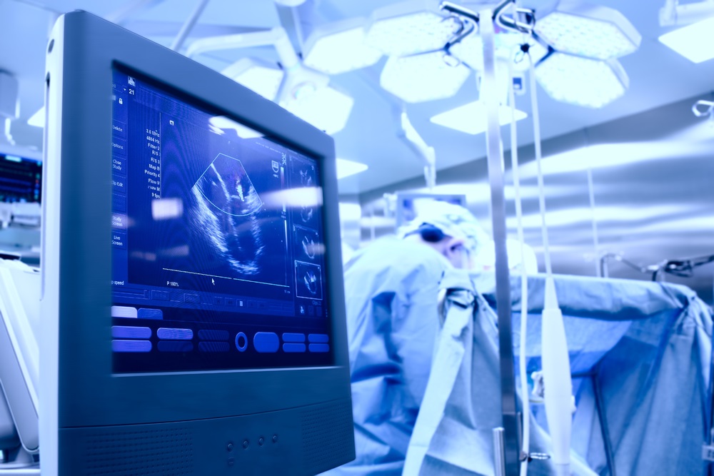
An echocardiogram is a test in which the human heart is examined using sound waves of a high frequency known as ultrasound technology. A cross section part of the heart in motion along with its chambers, major blood vessels exiting from the left and right ventricles and valves are displayed; also seen are factors such as the size of the heart chambers, the pumping functions, the valve functioning, the volume, the amount of fluid, presence of blood clots or tumors if any, within the heart, etc.
Thus echocardiography provides an action picture of the heart. Sound waves are sent through a device called the transducer through the human body, which return to the transducer as echoes after bounce off of the heart. These echoes transformed into images on a monitor in order to produce pictures of the heart. The echocardiography technology is either a one dimensional also known as “M-mode” echo cardiography wherein one beam of ultrasound is directed towards the heart to see only the left side or the primary pumping chamber of the heart or a two dimensional echo cardiography which provides a detailed picture of the heart.
 |
| Examination Tables |
Also used is the Doppler echocardiography in order to measure the quantum of blood flowing via the arteries producing a pattern of blood flow within the heart.
No cut to the skin
One of the important aspect to be borne in mind is that modern technology does offer several advantages as compared to the ones that were in practise hitherto. Diagnosing with the assistance of echocardiography ensures that there are no cuts or injury to the skin or any part of the human body yet delivering the results being sought from the test. Zero injury also means zero care to the skin from fungal infections and the like. An echocardiogram report is merely a report which gives the status of the heart, the valves and other components within the heart such as the ventricles, etc. using sound waves at high frequency – known as ultrasound waves.
The echo test shows moving images and takes pictures of the condition of the heart of a patient. These reports help the specialist evaluate the health of the heart and recommend treatment if necessary. An echo cardiogram specialist first helps the patient lie down on an echocardiography table. Enough gel is applied over the chest area which makes sliding a microphone-like device over the chest easy. This procedure provides a live picture of the heart and valves. Radiation is not involved in heart ultrasound thus there is no risk.
Table used for echocardiography
The table on which the procedure of echocardiography is carried out is the echocardiography table. This table has been created keeping the sonographer technician in mind and it helps in providing an imaging experience which is free of barriers of whatsoever nature. As a result best quality images are achieved and there is no injury to the sonographer technician. To carry out an effective test it is necessary for the sonographer technician to have access to the patient from all the sides of the table.
The cardiac scanning cushion is dropped down and can be released from both the sides of the table resulting in zero protruding. This gives full open access to the left thorax part to the sonographer for a non-obstructed approach and imaging. The side rails can be flushed to the table and folded fully under the table such that there is nothing in the working path of the sonographer and thus eliminating all possible obstruction and also preventing any injury either to the muscles or other parts of the patient's body. The top part of the table is made keeping the comfort of the patient in mind and this houses anti-microbial and anti-bacterial covers, ensuring full health protection against all types of bacteria and also ensuring a nice smoother feel making it very comfortable for the patient.
Additional options such as the extendable scanning arm with ability to rotate up to 120 degrees and a soft head rest extension provides easy thyroid and artery access makes the Echocardiography table indeed unique. The weight carrying capacity of the table is up to 300 kgs and can lift very heavy patients too without any problems. The table is also equipped with fully adjustable heights for easy transfer to a wheelchair should the need arise. The sleek base design ensures obstruction-free compatibility with all ultra sound equipments and access to the sonographer.
 |
| Echocardiography tables |
New Features
- Other options such as the scissor movement for the legs that form part of the frame of the table are also available which are controlled by means of a motor which rises and falls in slow and steady motion in accordance to the height requirements of the sonographer and the patient alike; rising to help the sonographer attend to the patient and fall to help the patient out of the table bed after the test is completed. Adjustment to the upper torso could be done in several ways namely, manual, hydraulic or height adjustment using electrical appliance. Height adjustment ranges from 450 mm to 950 mm as per the requirement.
- A sturdy frame manufactured with a dual box rectangle profile also helps avoid rocking movement or swinging of the table structure thus providing stability. Manual height adjustment is done with the help of a lever provided at one end of the table which could be pushed back after use so that it does not hinder the movement around the table. The Hydraulic movement is done using a foot pump located under the table on both sides of the table for ease of use by the sonographer. Electrical adjustment is controlled by ultra-silent motor and a hand switch which are adequately coated to guarantee maximum comfort for the user.
Hope you will get all the necessary information about echocardiography tables. Get in touch with us to get more information about examination tables.


0 comments